
Neurology and Spine
Craniofacial Reconstruction Surgery
Craniofacial surgery is used to treat issues with the head, skull, face, and neck. Specifically, the treatment involves surgical procedures and the supervision of skilled surgeons. Craniofacial reconstruction surgery reconstructs the damaged bone and tissue; as a result, this helps in improving the appearance of disfigured or abnormal shapes of the face and head. For instance, children with abnormal facial structures benefit from early surgery, which is advantageous in minimizing the impact of these conditions on growth, development, and function. -br There are generally two groups of people who may need craniofacial surgery. First, the first group of people are those who are born with abnormalities of the bones, muscles, and other tissues of the skull and face. This condition is known as congenital abnormalities. -br On the other hand, the second group includes people who develop irregularities in the skull and face later in life. This can occur due to diseases and trauma. This condition is known as acquired abnormalities.

Neurology and Spine
Discectomy
A discectomy is a surgical procedure to remove some part or complete part of an intervertebral disk in the spine. To explain further, intervertebral disks are like flat, round cushions that are placed between the vertebrae (bones) in the spine and work as spine shock absorbers. Each disk has a soft, gel-like centre called nucleus pulposus, surrounded by a flexible outer ring called annulus. Importantly, intervertebral disks are under regular pressure. As a result, a disk can tear, allowing some of the nucleus gel substances that come out through leaking. This condition is known as a herniated disk, also known as bulging, slipped, or ruptured disk. Consequently, this is the main reason that leads to performing a discectomy. -br In most cases, herniated disks mostly affect the lower back called the lumbar spine, but in addition, they also affect the neck called cervical spine too. However, a rare place where herniated disks are seen is the middle back. -br A discectomy in India, also spelled as diskectomy.

Neurology and Spine
Lumbar Decompression Surgery
Lumbar decompression surgery is a surgical intervention that is performed to treat compressed nerves in the lower spine. This surgery is performed only when other non-surgical treatments fail to relieve. The surgery is helpful in providing relief and other symptoms like numbness in the legs on the nerves in the spine.

Neurology and Spine
Meningioma
Meninges are the layer of tissue that protects the brain and spinal cord. A meningioma is a brain tumor that develops in meninges. Meninges are made of three layer tissues that cover and protect the brain and spinal cord. Meningiomas start from cells made up of particular arachnoid cells which are found in a thin, web-like layer that covers the brain and spinal cord. This layer is one of those layers that protects and makes up the brain's covering protective system known as meninges. -br Meningiomas in India are not always cancerous (benign), although sometimes they can be cancerous and malignant. If a tumor is cancerous it means it's aggressive or can spread out in other tissues as well or can spread in other parts of the body. A benign tumor is the type of tumor that can't spread to other parts of the body. -br Meningiomas are mostly located near the top or outer curve of the brain. They also form at the base of the skull. Spinal meningiomas are very uncommon. -br Meningiomas develop slowly and inward. They do not show any symptoms before they are diagnosed. Even benign meningiomas can grow larger enough to be life threatening if they compress and affect nearby areas of the brain.

Neurology and Spine
Spina Bifida
Spina bifida is a medical condition of an unborn fetus that means incomplete development of the spine during the first month of pregnancy. Moreover, this is the most common inborn disorder that is mostly affected in the United States. The term spina bifida means split spine. -br Specifically, spina bifida occurs during the first 28 days of pregnancy, even before the pregnancy is confirmed by the women. The condition is a type of neural tube defect (NTD). -br In addition, in some uncommon cases, the spina bifida in India is very serious. The condition varies widely in degree. Generally, most of the cases are mild that have no symptoms and even can be treated without any serious treatment. -br However, in more uncommon cases of such disorder, infants are born with open lesions on their spine that lead to damage to surrounding nerves and spinal cord. The opening can be treated through surgery, but the nerve damage can't be solved and results in permanent disability. Furthermore, spina bifida can take place anywhere in the back bone.

Neurology and Spine
Spinal Fusion Surgery
Spinal Fusion Surgery involves joining two or more vertebrae to eliminate motion between them, providing stability and reducing pain from spinal instability or deformity. -br The procedure can treat conditions like spondylolisthesis, degenerative disc disease, or fractures. Bone grafts and implants such as rods or screws are used to facilitate fusion and long-term spinal alignment.

Neurology and Spine
Cerebral of Brain Aneurysm
Cerebral aneurysm, also known as brain aneurysm, is a small and thin spot on the artery in the brain that bulges out and fills with blood. As a result, the bulging aneurysm leads to force or puts pressure on the nerves or brain tissue. This can also result in a burst, spilling blood surrounding tissue, known as hemorrhage. Consequently, a ruptured aneurysm can be a life-threatening condition, such as hemorrhagic stroke, brain damage, coma, or sometimes death.-br However, some cerebral aneurysms do not bleed, and are very small in size or cause other complications. These types of aneurysms are diagnosed while imaging tests for different medical problems. Cerebral aneurysms can take place anywhere in the brain, but most develop in major arteries along the base of the skull.-br Cerebral of brain aneurysms can develop at any age and anywhere in the brain. They are, however, most common in adults between the ages of 30 and 60 years, and are more common in females than males. Additionally, some patients are at high risk due to inherited disorders.

Neurology and Spine
Deep Brain Stimulation for Parkinson's Disease
Deep brain stimulation is a technique and a medical procedure that proceeds with a mild electric current delivered to the specific part of the brain. This means that this electricity in the current stimulates the brain cells in the area that treats several conditions of the brain and body. Specifically, the current travels through one wire or more wires and reaches to the brain connected with a small device implanted underneath the skin near the collarbone. -br Currently, deep brain stimulation is used for conditions like Parkinson's disease and epilepsy, although research is still on if it can be helpful for other conditions too. -br In this procedure, the treatment involves an implanted device that delivers electrical current directly to the brain areas.

Neurology and Spine
Early Onset Scoliosis Surgery
Early onset scoliosis involves the development of abnormal lateral curvature of the spine before the age of 10. The condition occurs at congenital or acquired different challenges because the spine is still in a growing stage. The majority of the children are diagnosed with this condition between the ages of 10 to 15 with adolescent idiopathic scoliosis. Identifying idiopathic scoliosis at birth or in early childhood is rare. The type of scoliosis is categorized by age at onset of diagnosis:

Neurology and Spine
Neurolysis Procedure
Neurolysis is a procedure that consists of chemical injection that involves pain relief management. Generally, this is typically for nerve pain, cancer pain or visceral pain. Depending on the case, different types of chemical neurolysis procedure based on which nerve it's pointing out, such as celiac plexus neurolysis and intercostal nerve neurolysis. Typically, the injected chemical consists of alcohol, phenol or glycerol. -br To understand this, nerves are like the wires of the body that travels the electrical signal between the brain and the body. These electrical signals lead to feeling sensation like touch, pinch and pain and move the muscles. Therefore, the goal of neurolysis is to stop the targeted nerve from sending pain signals to the brain. As a result, if these nerves stop sending signals, the body won't be able to feel any sensation or do not respond accordingly. -br In general terms, intentional destruction of the pain management through the nerves is said to be as neurolysis in general ways. In other words, this neurolysis in India is a term used to describe the work of the nerve that passes the signals to the brain.

Neurology and Spine
Spinal Cord Stimulator
A spinal cord stimulator is a device that is used to manage intense pain, a spinal cord stimulator is an implanted device. Specifically, the device is implanted in the body and controlled by the patient. In general, these devices are helpful in providing relief from various conditions. A spinal cord stimulator involves a thin wire that is called electrodes as well as a small pacemaker-like battery pack known as a generator. A spinal cord stimulator allows patients to use remote control whenever they feel pain; the device sends electrical signals. -br Importantly, these devices are placed with very own professional physicians, and that too with highly specialized training in interventional pain management under X-ray and ultrasound guidance.

Neurology and Spine
Anterior Cervical Discectomy & Fusion
Anterior cervical discectomy and fusion (ACDF) is a surgical procedure that treats pain caused by compressed nerves in the neck. This surgical approach involves removing the disk between the affected bones and fusing them together. In turn, ACDF surgery moves the pressure on the nerves or spinal cord. This procedure treats the issue in the neck through the front or throat area. Additionally, this procedure also helps in treating different types of arthritis that cause pain. -br ACDF in India surgery removes the pressure from the nerves around the spinal cord. The reason for the pressure is due to bone spurs or bulging disks. Bone spurs form as a result of arthritis. This disc performs as a shock absorber between vertebrae that slips out of place and moves up against a nerve. -br However, ACDF surgery can lead to weakness or damage to the spinal cord. These problems may occur due to accidents or with other serious problems. The pressure on the spinal nerves or spinal cord develops gradually.

Neurology and Spine
Corpus Callosotomy
Corpus Callosotomy is a surgical procedure to treat epilepsy. Epilepsy, which is a chronic medical condition, affects individuals for the long term and leads to recurrent seizures in both children and adults. Typically, the treatment is performed under the care of the best neurosurgeons in India. During the corpus callosotomy procedure, an incision is made through the brain's corpus callosum. The corpus callosum, which is a type of band of nerve fibers, joins the two halves of the brain (hemispheres) and relays messages from one hemisphere to the other. As a result, the corpus callosum prevents seizure signals from crossing back and forth between the two hemispheres, thus limiting the spread of seizure activity. This surgery is also known as callosal sectioning or brain splitting. -br Epilepsy surgery consists of various types, and corpus callosotomy is one of them. However, corpus callosotomy surgery is only recommended when antiseizure medications fail to relieve or treat the seizures effectively. In general, the entire procedure is performed under the supervision of experienced surgeons, involving a careful surgical cut of the band of fibers that join the two halves of the brain.

Neurology and Spine
Spinal Cord Injury- Stem Cell
Spinal cord injury (SCI) is caused due to traumatic injury, such as vehicle accidents, falling from the height, and sports injuries. Moreover, the seriousness of the injury depends on the location and how much it is injured, which sometimes include paralysis, loss of sensation, and dysfunction of motor skills below the level of injury. As a result, patients sometimes feel difficulties with bladder and bowel control, sexual dysfunction, and chronic pain that, in turn, has a severe impact on the quality of life and the person's individuality.

Neurology and Spine
Spinal Dysraphism
Spinal Dysraphism refers to congenital malformations of the spinal cord and vertebrae, such as spina bifida or tethered cord. -br Surgery aims to correct structural abnormalities, relieve tension on neural tissues, and prevent progressive neurological damage. Early intervention improves functional outcomes and quality of life.

Neurology and Spine
Spinal Stenosis
Spinal Stenosis refers to the narrowing of the spinal canal, which can compress nerves and lead to pain, numbness, or weakness. -br Surgery involves decompressing the spinal cord or nerves to relieve symptoms and restore function. Timely intervention prevents progressive neurological damage and improves quality of life.

Neurology and Spine
VP Shunting
VP Shunting involves placing a catheter from the brain’s ventricles to the peritoneal cavity to drain excess cerebrospinal fluid (CSF), relieving pressure. -br This procedure is commonly performed in hydrocephalus, where fluid accumulation leads to neurological symptoms. VP shunts are adjustable and can provide long-term CSF management.

Neurology and Spine
Spinal Decompression Surgery
Spinal decompression surgery is a surgical procedure performed to relieve symptoms that are associated with compression of the spinal cord and of its origin. These may include back or neck pain and also radiating limb pain. Moreover, the relieving of pressure on the spinal cord or nerves is the main motive of spinal decompression surgery, this occurs due to herniated disc, spinal stenosis and spondylolisthesis. In addition, the method is helpful in relieving pain and improves mobility. Furthermore, it also means to reduce pain, improves the quality of life, and improves the issues related to back pain that is experienced on a daily basis. On the other hand, spinal decompression in India also provides non-surgical options to alleviate pressure on the spinal structure. Consequently, this is more beneficial in creating more space within the spinal canal, reduces merge compression, and related symptoms.

Neurology and Spine
Cerebral Angioplasty
Cerebral angioplasty is a procedure that treats stenosis (narrowing) or complete occlusion (blockage) of the blood vessel in the brain. As a result, this procedure results in reducing blood flow and oxygen supply. Consequently, this can lead to strokes or other neurological issues. Cerebral angioplasty in India is a skilled endovascular procedure performed by a neurointerventional radiologist or interventional neuroradiologists. In simple words, cerebral angioplasty is a minimally invasive procedure that is used to treat blockage in the blood vessels of the brain. Specifically, this technique is performed with a catheter, a thin tube, used to access and repair narrow or blocked vessels, therefore this also restores proper blood flow to the brain.

Neurology and Spine
Minimally Invasive Spine Surgery
Minimally invasive spine surgery is a method to view and access the spine using the technique that does not damage the tissue and nearby muscles. In contrast, in a traditional method of open surgery, the surgeon makes a long incision through the skin. To explain further, to get access to the site, the surgeon makes a large incision and a large amount of muscle and nearby soft tissue are pulled away from the bone. As a result, this leads to increased recovery time and is more painful after the surgery. -br On the other hand, the other method, which is minimally invasive surgery, is more advanced techniques that need small and few incisions through the skin. In this method, a thin tube or endoscope passes through the cut that is made by the surgeon allowing it to work through a small incision area. Consequently, this results in better ways like less damage to the muscle,soft tissue, and skin. Moreover, this also requires less time to recover and is also helpful in faster recovery.

Neurology and Spine
Transforaminal Lumbar Interbody Fusion-TLIF
Transforaminal Lumbar Interbody Fusion (TLIF) is a minimally invasive spinal fusion technique that stabilizes the lumbar spine by placing bone grafts and implants through a posterior approach. -br This procedure relieves pain caused by degenerative disc disease, spondylolisthesis, or spinal instability, while maintaining alignment and preserving neural structures.

Neurology and Spine
Arteriovenous Malformations
Arteriovenous malformations occur when a group of blood vessels develops incorrectly in the body. In these cases, veins and arteries are jumbled together and form a direct connection, bypassing normal tissues. This condition occurs during development when the child is in the womb or shortly after birth. -br Typically, this doesn't show any symptoms or issues. Instead, this problem shows up once the patient is having some other unrelated health issues. For instance, a single ruptured blood vessel in an AVM leads to the issue that needs medical attention. Moreover, in some cases, AVMs are found during autopsy after death. -br An abnormal tangle of blood vessels that normally looks like a web. This tangle is made of arteries that circulate blood to the brain and veins that normally drain blood from brain tissue. These arteries carry oxygen-rich blood from the heart to the brain and also to the other organs and tissues. Then, again, veins supply oxygen- and nutrient-poor blood and waste material from tissue to the heart and lungs. -br This exchange process occurs in capillaries, which is the place where the smallest blood vessels of arteries and veins connect. As a result, this can also be a reason for high-flow arterial blood to connect directly to veins. Ultimately, this abnormal connection between artery and vein in an AVM can lead to rupture of the vessel and bleeding into the brain.

Neurology and Spine
Brachial Plexus Surgery
The brachial plexus works to convey movement and sensory signals from the upper spinal cord in the neck down into the arms and hands. Essentially, this is a network of nerves that is useful to convey signals. However, an accident or trauma to the neck or shoulder can injure the brachial plexus, which also causes pain, weakness, numbness, or paralysis in the arms or hands. -br In some cases, some brachial plexus injuries are able to cure on their own and restore normal or near-normal function. On the other hand, in the case of a severe injury, brachial plexus surgery is considered to be the best treatment to relieve the symptoms of pain and restore sensation and mobility.

Neurology and Spine
Skull Base Surgery
Skull base surgery is performed to remove cancer and non-cancer growths or the abnormal development inside the brain, the skull base, or the vertebrae of the spinal column. This is because the area is hard to see and reach. The skull is made up of bones and cartilage that develop the face and cranium, which surrounds the brain. In fact, the bone of the skull can easily be felt by touch on the top of the skull. The 5 bones that form the bottom base of the cranium also form the eye socket, top of the nasal cavity, some of the sinuses, and the bones around the inner ear. Moreover, the skull base is a complex and sensitive area that has various openings. Through these openings, many blood vessels, spinal cord, and nerves pass. -br Skull base surgery in India can be done through endoscopic procedure; therefore, it comes under a minimally invasive procedure. In this method, the surgeon inserts a special tool through the nose or through the mouth, or alternatively, the surgeon makes the hole above the eyebrow and inserts the tool from there. Importantly, this type of surgery is done under the supervision of skilled surgeons and a team of specialists.

Neurology and Spine
Sacral Nerve Stimulation-SNS
Sacral nerve stimulation (SNS) is a procedure that helps the patient with problems of bowel and bladder. Moreover, sacral neuromodulation therapy helps to improve and restore normal bladder or bowel function. Specifically, sacral nerve stimulation (SNS) in India uses an implanted device that sends mild electric impulses to stimulate nerves that control the function of bowel and bladder. These nerves, in turn, help to manage the function of peep and poop. Consequently, the method changes the signals between the brain, spinal cord, bowel and bladder. This function, therefore, is known as sacral neuromodulation therapy. -br Urinary and fecal incontinence has a severe impact on the life of a person. Sometimes it can be the reason for discomfort, embarrassment or it can increase the dependency on others. Patients who have no result with conventional therapies, SNS plays an important role for such treatments.

Neurology and Spine
Spine Tumor Surgery
Spine Tumor Surgery involves removing primary or metastatic tumors from the spinal column to prevent neurological deficits, relieve pain, and restore stability. -br Careful preoperative planning, imaging, and collaboration with oncology specialists ensure safe resection while preserving spinal cord function.

Neurology and Spine
Artificial Spine Lumbar Disc Replacement
An artificial spine lumbar disk replacement is a surgical procedure to remove and replace the damaged or worn disk material between the small bones in the spine (vertebrae). The replacement occurs with an artificial prosthetic or artificial disk. In essence, the main aim of the removal and replacement is to relieve pain symptoms while maintaining the normal motion and function. However, it is allowed with some other procedures, such as spinal fusion. For instance, the approximate estimate is that 70 to 80% of people will experience low back pain at some point in their lives; nevertheless, some of them can improve with non-surgical treatments. -br Surgery is only recommended when other non-surgical treatments fail to relieve symptoms. Typically, pain should be caused by one or two worn-out (arthritic) discs, which can be diagnosed through tests and a physical check-up. Moreover, when the patient has already gone through fusion surgery that heals perfectly but ends up with no pain improvement, further evaluation is needed. -br The belief of failure to improve after fusion surgery is due to the reason that fusion prevents normal motion in the spine. Artificial disc replacement helps to preserve normal motion that leads to perfect treatment for low back pain.

Neurology and Spine
Brain Biopsy
A brain biopsy is a method to diagnose brain illness. The procedure, in fact, involves the removal of a tumor or a piece of tissue from the brain to examine under a microscope. The main types of brain biopsy consist of:

Neurology and Spine
Embolization of Brain & Spinal Tumors
Embolization of the brain and spinal tumor is a technique that blocks the blood vessels artificially, that hold or stop the blood flow to the tumor. As a result, this leads to the tumor that stops supply of blood and oxygen and also results in the slow growth of the tumor or sometimes it is destroyed in the same way. Specifically, tumors in the head and neck along with the spine are usually treated by blocking the blood supply of the tumor by using a special liquid. This procedure is carried out by inserting a thin tube called catheters through the groin or arm and guiding them through the way of blood vessels to the tumor. Alternatively, there is one option more and that is to perform the procedure by going straight through the skin to reach the tumor.

Neurology and Spine
Foraminotomy
A foraminotomy is a surgical intervention performed to relieve pressure on the nerve roots in the spine. Specifically, the procedure is used to open up the foramen or open the vertebra bone, a place where the nerve roots exit the spinal canal. In fact, the peripheral nervous system makes up the foramen, the nerve that exits the spinal canal. In case the foramen is narrow, the person can feel the nerve pain and other symptoms that may affect the movement. Therefore, a foraminotomy can be beneficial in relieving pressure and reducing pain. -br Furthermore, a foraminotomy in India is a surgical procedure that enlarges the area around one of the compressed nerves in the spinal column. The spine consists of a chain of bones known as vertebrae. The intervertebral disks are placed between vertebrae and work as a cushion. -br Sometimes, the compressed nerve can cause symptoms such as pain, tingling in the arms and legs, or weakness. Ultimately, the accurate symptoms depend on the location of the compressed nerve along the spinal column.

Neurology and Spine
Vertebroplasty & Kyphoplasty
Vertebroplasty and Kyphoplasty are minimally invasive spine procedures designed to treat vertebral compression fractures, often caused by osteoporosis or trauma. -br Vertebroplasty involves injecting bone cement directly into the fractured vertebra to stabilize it. Kyphoplasty includes the use of a balloon to restore vertebral height before cement injection, which can also help correct spinal deformity.

Neurology and Spine
Anterior Cervical Corpectomy
An anterior cervical corpectomy is a surgical intervention performed to remove the damaged vertebrae and intervertebral disc from the spine in the neck. In particular, a corpectomy is a procedure that removes damaged vertebrae and intervertebral discs that are crushing the spinal cord and spinal nerves. Anterior means the front of the body, while cervical means the spine in the neck.-br Moreover, a corpectomy is almost similar to a procedure called discectomy, as both procedures are performed to treat similar conditions. However, the main difference is that corpectomy is a more complex and vast procedure. This is performed when the disease enlarges beyond the areas that can't be treated with discectomy. Discectomy stands for removing the damaged part of the disc and bone spurs, whereas, in corpectomy, the disc, bone, spurs, and vertebrae are removed.

Neurology and Spine
Blood Clot Brain Surgery
Blood clots are formed when the inside of blood vessels obstructs the blood flow. This is generally a natural response in the healing process that prevents bleeding. However, this can be a life-threatening condition if not treated. -br A blood clot in the brain develops when the blood vessel becomes obstructed, blocked, or ruptured, which disrupts the flow of oxygen and nutrients in the brain. Consequently, this can be severe in the case of damage to brain cells, stroke, or sometimes death. Moreover, this can cause serious complications if not noticed, such as seizures or paralysis. On the other hand, while some patients experience mild symptoms, it is important to understand the risks and symptoms of blood clots, as they are essential to require medical treatment. Therefore, consult a doctor if you notice any signs or symptoms, as it is crucial to seek treatment promptly.

Neurology and Spine
Brain Tumour Surgery
Surgery is one of the most common and main treatments for patients with brain tumors and spinal cord tumors. The surgery is recommended for:

Neurology and Spine
Microvascular Decompression-MVD
Microvascular decompression (MVD) is a surgical intervention performed to relieve abnormal compression of a cranial nerve causing trigeminal neuralgia, glossopharyngeal neuralgia, or hemifacial spasm. Specifically, MVD in India is a surgery that is performed to relieve symptoms of pain and muscle twitching caused due to nerve compression by an artery or vein. To achieve this, the surgery procedure involves the direct access of the skull by opening and exposing the nerve at the base of the brainstem to insert a small sponge between the compressing vessel and the nerve. In this process, the small incision or cut is made behind the ear. Then, while viewing the trigeminal nerve through a microscope, the surgeon places a soft cushion between the nerve and injured blood vessels. Ultimately, the main goal of MVD is to identify and relieve pressure from one or more blood vessels. This step is crucial in finding blood vessels pressing on the trigeminal nerve and gently shifting them away. Finally, a small cushion is placed between them to prevent the pressure and relieve pain.

Neurology and Spine
Spinal Disc Replacement
Spinal Disc Replacement involves replacing a damaged intervertebral disc with an artificial implant to restore spinal mobility and reduce pain. -br Unlike fusion, disc replacement maintains motion at the affected level, potentially reducing stress on adjacent vertebrae and minimizing long-term degeneration.

Neurology and Spine
Spinal Endoscopic Surgery
Spinal Endoscopic Surgery uses small incisions and a camera-equipped endoscope to treat spinal conditions like herniated discs or stenosis. -br This minimally invasive technique reduces tissue damage, shortens recovery time, and often allows faster return to daily activities compared to traditional open surgery.

Neurology and Spine
Comprehensive Myelopathy - Spinal Cord Surgery
Myelopathy is a spinal cord compression, or an injury to the spinal cord. As a result, it can lead to pain, numbness, and problems in moving particular parts of the body. Myelopathy occurs due to traumatic injury, aging, or herniated disk. In fact, myelopathy symptoms can be severe and worsen if left untreated or ignored. -br Myelopathy in India is a group of symptoms caused by spinal cord compression or an injured spinal cord. When some pressure or force compresses the spinal cord, it becomes difficult to move properly. Consequently, this can lead to pain, loss of feeling, or problems in moving certain parts of the body. -br The spinal cord is a collection of nerves that travels messages between the brain and body. It is placed inside the hollow area of the spine. Furthermore, the bone in the spine acts as a shield of protection for the spinal cord. Vertebrae can become weak due to several diseases and conditions. -br For instance, the loss of feeling or a feeling of pins and needles in the hand after leaning on the elbow for too long may occur. However, this condition can become worse if it remains untreated.

Neurology and Spine
CyberKnife
The cyberKnife is an advanced form of radio robotic surgery. To begin with, radiosurgery is a treatment that involves the exact and concentrated doses of radiation therapy to specific areas of the body. Importantly, this surgery is done without any incisions or anesthesia, also known as noninvasive treatment. Moreover, cyberKnife is very much accurate, so there is a minimal exposure to surrounding healthy tissues while performing the treatment. -br In terms of usage, the cyberKnife is used to treat tumors—both types of tumors, cancerous or non-cancerous. Generally, this treatment is only recommended if the condition is hard to treat with surgery. -br In addition, cyberKnife treatment is painless, non-surgical, and precise, targeting radiation to destroy tumors or lesions within the specific area of the body. -br Lastly, the cyberKnife is flexible with robotic arms that help in the treatment of areas like the spine and spinal cord, which are impossible to treat by other techniques.
.png&w=256&q=75)
Neurology and Spine
Kyphosis (hunchback)
Kyphosis (hunchback) is an abnormal shape of the spine which, as a result, is curved outward more than it should be. In contrast, a normal spine is straight in shape when viewed from behind. Consequently, this causes the upper back around the thoracic region to be affected. The thoracic region is a portion of the spine between your neck and ribs. -br Visually, the abnormal shape of the spine looks like a rounded or humpback. Kyphosis in India is usually defined as a curvature of the spine that measures 50 degrees or more on an X-ray. Furthermore, some diagnostic tests use invisible electromagnetic energy beams to release images of internal tissues, bones, and organs onto film. Typically, the normal spine bends from 20 to 45 degrees of curvature in the upper back area.

Neurology and Spine
Carotid Endarterectomy
A carotid endarterectomy is a surgical procedure that involves the treatment of carotid artery disease. The carotid arteries, in fact, are one of the main blood vessels that transfer oxygen and blood to the brain. When these arteries become narrowed, it reduces the blood flow to the brain, which can eventually lead to a stroke. -br During a carotid endarterectomy, using a surgical approach, the surgeon removes plaque that has formed inside the carotid artery. This is done by making an incision on the side of the neck over the affected carotid artery. After opening the artery, the plaque is carefully removed. Once the plaque is removed, the artery is stitched back as before. This procedure helps restore the normal flow of blood to the brain. -br It is important to note that this procedure is performed under general anesthesia, although the patient is not completely in a sleep state.

Neurology and Spine
Cranioplasty
Cranioplasty is a surgical procedure that treats or repairs a defect in the skull. Skull is one of the most important and protective parts of the brain. The skull is the bone that surrounds and protects the brain. Cranioplasty helps to repair a defect in the skull that gives protection to the brain from getting damaged. Cranioplasty surgery is recommended after certain forms of brain surgery or any traumatic injury. The main aim of the cranioplasty procedure is to repair and reshape a defect in the skull. -br During the surgical intervention, the surgeon may perform:

Neurology and Spine
Temporal Lobectomy
Temporal Lobectomy involves removing a portion of the temporal lobe to control seizures in patients with refractory epilepsy. -br This surgery is considered when medication fails to prevent frequent or severe seizures. Advanced imaging and EEG monitoring help identify the seizure focus before surgery.

Neurology and Spine
Ventriculostomy
Ventriculostomy is a surgical procedure that involves creating a small opening in the brain’s ventricular system to drain excess cerebrospinal fluid (CSF). -br It is commonly performed to treat hydrocephalus, elevated intracranial pressure, or to administer medication directly into the ventricles. The procedure provides rapid relief and can be life-saving in emergencies.

Neurology and Spine
Craniotomy Surgery
A craniotomy is a surgery that gives access to the brain; in fact, the surgery is performed to remove a piece of skull to get proper access to the brain. Furthermore, the surgery is performed under the top neurosurgeon in India, and during the same surgery, the surgeon re-applies the piece of skull. -br In addition, a craniotomy is a major surgery. Moreover, this type of condition is usually diagnosed during the treatment of a brain tumor or some type of traumatic brain injury.

Neurology and Spine
Kyphoplasty
Kyphoplasty is a minimally invasive procedure to treat compression fractures in the spine. To clarify, compression fracture is a break or cracks in the vertebrae. The vertebral body is a thick rounded part on the front of each vertebrae. When a fracture occurs in the spine, it leads to weakening or collapsing the spine. Moreover, these fractures can also affect the posture in the future. In fact, compression fractures can lead the bones in the spine to collapse, resulting in kyphosis or curve in the spine. Consequently, this can be an obstruction in breathing or can limit the function of the abdominal organs. Therefore, kyphoplasty helps to stop this curve from occurring. Additionally, sometimes kyphoplasty is referred to as balloon kyphoplasty by the healthcare professionals.

Neurology and Spine
Peripheral Nerve Surgery
Peripheral nerve surgery is a surgical procedure that improves the function and reduces pain and disability in patients with peripheral nerve disorders. For example, some of the disorders are acute nerve injury, entrapment neuropathies, and nerve sheath tumors. In such cases, peripheral nerve injury helps to turn healthy nerves to take over the function of the nerves affected by the injury, disease or other conditions. -br To explain further, the peripheral nerve is the route that joins the brain and spinal cord to other parts of the body. As a result, peripheral nerve disorders result in preventing messages from the brain to the rest of the body and also can impact one nerve or many nerves. -br A team of healthcare professionals that lead to performing the surgical treatment as well as rehabilitation include:

Neurology and Spine
Spinal Instrumentation
Spinal Instrumentation involves using medical implants such as rods, screws, plates, and cages to stabilize the spine during corrective or fusion procedures. -br These devices provide structural support, restore alignment, and protect neural structures during healing. Instrumentation is commonly combined with fusion or deformity correction surgeries.

Neurology and Spine
Acoustic Neuroma
An acoustic neuroma is a non-cancerous, benign ear tumor that is called vestibular schwannoma. It affects the hearing and sensing balance. In addition, the tumor affects the nerve that sends signals from the inner ear to the brain. This develops on the eighth cranial nerve. As a result, due to affecting the nerve, the brain does not receive signals to react and cannot hear and maintain the balance in between. Acoustic neuromas don't spread like cancerous tumors, however, when they grow, they affect hearing, causing some kind of sound like tinnitus. Moreover, it can lead to facial numbness and facial weakness. The treatment for this tumor is surgery, radiation therapy, and in uncommon cases, chemotherapy is also recommended for the tumor.

Neurology and Spine
Cervical Spine Surgery
Cervical spine surgery is performed to treat problems in the neck such as misalignment, compressed nerves, or spinal cord abnormalities. Some of the most common reasons to perform cervical spine surgery include:

Neurology and Spine
Endovascular Coiling
Endovascular coiling is used to block blood flow into an aneurysm. An aneurysm, in other words, is a weakened area in the wall of an artery. This procedure, also known as endovascular embolization, is performed to address this issue. In case the aneurysm ruptures or gets broken, it comes leading to a life-threatening condition due to bleeding and brain damage. Therefore, preventing the flow of blood into an aneurysm helps to keep it from breaking or rupturing. -br To perform the procedure, healthcare professionals use a thin tube called a catheter into a groin artery for endovascular coiling. From there, the catheter moves into the affected brain artery and then puts the coil in place. X-ray imaging helps to guide the catheter into the artery. The coils are made of soft platinum metal and are shaped like a spring. Additionally, the coils are very small and thin in shape. Sometimes, the coils are used with stents to stop the coils from moving out of the aneurysm. -br Moreover, endovascular coiling in India is used to treat conditions called arteriovenous malformations. An AVM is a tangle of blood vessels that causes an abnormal link between an artery and a vein. Notably, this can take place anywhere in the brain or spinal cord.

Neurology and Spine
Lumbar Disc Microsurgery
Lumbar disc microsurgery, also known as lumbar microdiscectomy, is a surgical intervention that performs to remove a piece of intervertebral disc that is pressing on the spinal nerve. It can lead to severe pain, weakness, or numbness in the lumbar or lower back. Consequently, the pain may spread downward to the length of the leg, and it is called radicular pain. Furthermore, the largest moveable segment of the vertebral column is the lumbar that is also at risk of painful disorders. This is because the spine is affected by twisting and bending. Moreover, this is also meant to bear the most of the weight of the body.

Neurology and Spine
Microdiscectomy
Microdiscectomy, also known as microdecompression or microdiskectomy, is a minimally invasive surgical procedure performed to treat the patient having herniated lumbar disc. India is among the nation's providing advanced treatment of earlier spinal surgery once known for larger incisions, long recovery and painful rehab. But yes, surgical advancement in the medical field has grown so far with immense technology and methods. Advancements like microdiscectomy in India procedures have improved the technique. -br During the microdiscectomy surgery, the surgeon will remove the small part of the herniated disc to relieve pressure on the spinal nerve column. -br Microdiscectomy is a surgical intervention that relieves pain symptoms and other problems caused when a herniated disc in the spine presses on an adjacent nerve root. During the surgery the nerve is released by removing small fragments of disc, bone and ligaments. -br Microdiscectomy is a minimally invasive spine that is performed with less and small incision and the surgeon used a microscope or surgical glasses called loupes to magnify and get the access to the site where the injury occurred.

Neurology and Spine
Pineal Region Tumors
Pineal region tumors are brain tumors that are found very rarely. Just behind the brain stem, the pineal gland is formed in the middle of the brain. The start in the pineal gland or tissues around it. It makes the hormone melatonin that controls sleep. The tumors in the area of the brain, the brain can obstruct or block the cerebrospinal fluid. The cerebrospinal fluid is a fluid that surrounds and protects the brain and the spinal cord. -br This can lead to block and build the cerebrospinal fluid in a specific area of the brain. This can lead to increased pressure in the brain. Some common symptoms of increased pressure include headache and feeling ill.

Neurology and Spine
Radiofrequency Rhizotomy- Neurotomy
Rhizotomy is a minimally invasive procedure that is performed to kill the nerve fibres and remove painful nerves that send pain signals to the brain. The nerve fibres can be finished and destroyed by cutting off with a surgical instrument or burning them with a chemical or electrical current. Rhizotomy is proven to give instant relief from pain that can be long lasting for years until the nerves recover and are able to send pain signals again. -br Rhizotomy- neurotomy also known as ablation or neurotomy, these terms are used to describe the removal or destroying of tissue that is causing pain.
Neurology and Spine
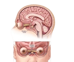
Pituitary Tumor
Pituitary tumors are the tumors that develop unusually in the pituitary gland. This gland is a type of pie in size. The location of the gland is behind the nose at the base of the brain. Due to tumors these pituitary glands develop an excessive amount of certain hormones that controls the important function of the body. -br Most of the tumors of the pituitary are benign. This means they are not cancerous. Another name for these pituitary non cancerous tumors is pituitary adenomas. Most adenomas stay in the pituitary gland or in the tissue around it, these tumors grow slowly and also don't spread to the other parts of the body. -br Pituitary tumors in India have several treatments. The tumors may be removed through surgical intervention. Depending upon its growth, if it's slow and less it can be controlled with medication or radiation therapy. Sometimes the hormone level may be controlled with medicine.

Neurology and Spine
Spinal Deformity Correction Surgery
Spinal Deformity Correction Surgery is performed to correct abnormal curvatures of the spine such as scoliosis or kyphosis. -br The procedure involves realigning the vertebrae, often using rods, screws, and other instrumentation, to restore proper posture and prevent progression of the deformity. Early intervention improves long-term functional outcomes.

Neurology and Spine
Gliomas
A glioma is a tumor that develops in the brain and spinal cord when the glial cells grow out of control. These cells support nerves and help the central nervous system. Gliomas usually grow in the brain and spinal cord. -br Gliomas are cancerous and grow slowly. They are primary brain tumors, these cells originate in the brain tissue. Gliomas don't usually spread outside of the brain or spine. These gliomas can be life threatening because they can:

Neurology and Spine
Lumbar Laminectomy for Spine
A laminectomy is a surgical procedure performed on the spine to remove the lamina. The bony arch or lamina helps to support and protect the back part of the spinal cord on the vertebrae. Removal of lamina relieves pressure on the nerves and spinal cord by making more space on the spinal cord. -br A laminectomy is performed in the lower spine that is lumbar laminectomy for spine, this is helpful in identifying the cause of nerve pain. This procedure is helpful in treating pain that is developed in the neck or in the middle of the back.

Neurology and Spine
Stereotactic Radiosurgery
Stereotactic Radiosurgery delivers highly focused radiation beams to treat brain tumors, arteriovenous malformations, or other small neurological lesions. -br The technique avoids open surgery, minimizing trauma while accurately targeting abnormal tissue. It is often used when traditional surgery carries higher risk or is not feasible.

Neurology and Spine
Anterior Cervical Discectomy
An anterior cervical discectomy is performed, along with the fixation and fusion, to check the stability of the spine. The discectomy procedure, in particular, involves the removal of a damaged, herniated intervertebral disc (the cushion between the bones of the spine) and arthritis. Furthermore, anterior surgical intervention stands for an approach from the front side of the body and cervical spine in the neck. In simple words, an anterior cervical discectomy is a surgical procedure that involves removing a damaged intervertebral disc from the spine in the neck, and that too uses a surgical intervention from the front of the neck.-brMoreover, a fusion is the implantation of the bone graft that fuses with vertebrae and grows with the bones. On the other hand, a fixation is the implantation of screws, rods, or plates. Fixation is used to fix the bones in position while they fuse. In some cases, an artificial disc or arthroplasty is used in place of fusion when the damaged disc has been removed. Patients with normal function of the cervical spine are, therefore, the most deserving patients for cervical artificial disc replacement.-brIn addition, an anterior cervical discectomy is a surgical procedure that is similar to a surgical procedure called anterior cervical corpectomy. However, the difference between them is that a corpectomy is a more extensive procedure used when the disease extends; it is usually performed when the disease affects areas that cannot be addressed by a discectomy alone.

Neurology and Spine
Robotic Brain Surgeries
Robotic brain surgery is a great innovation of the medical field. It represents an excellent advancement in the field of neurosurgery. Also it improves the safety and reduces the risk factors with great procedure and technology. This innovation involves robotic systems to assist the surgeons in showcasing the anatomy of the brain, allowing minimally invasive procedures. Robotic brain surgery in India has transformed and enhanced the traditional methods, resulting in the patient's successful recovery, taking less time to recover and greater precision with improved surgical capabilities.

Neurology and Spine
Scoliosis Spine Surgery
Scoliosis spine surgery is a condition where the spine is tilted, rotated or curved sideways in S or C shape. Then treatment to make the shape in a normal position is performed with scoliosis surgery. The curvature in the spine is more than 10 degrees and can be caused due to genetics, hormones, neurological issues, muscle diseases or overuse of the spine. Scoliosis affects the balance of the body, the body may tilt to one side, also may lead to breathing and neurological problems that impact the quality of life. Scoliosis surgery aims to stop curvature in the spine, straighten the spine and create stability to balance the spine.

Neurology and Spine
Spinal Decompression & Fusion
To start with, spine fusion surgery is a procedure that connects two or more vertebrae in the spine permanently and also stops movement to make the spine stabilize. In general, spinal fusion surgery aims to heal the process of broken bones. -br Moreover, fusion surgery is one of the best treatments for the patient with serious spondylolisthesis, degenerative disc disease or nerve compression with low back pain. In some cases, the instability in the spine can be due to traumatic injury or accident. Additionally, another cause might be the spinal decompression procedure that has created spinal instability by removing bone, disc, soft tissue that needs a spinal fusion surgery. -br Typically, surgical intervention includes instrument and fusion. Specifically, instrumentation leads to devices such as cages, metal plates, screws, and rods that are useful in holding the vertebrae together so that it heals and becomes solid together. Meanwhile, fusion involves autograft or allograft that is covered into or around instrumentation.

