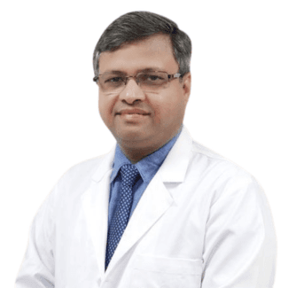

Dr. Nagesh Chandra

Neurology and Spine

Dr. Nagesh Chandra

Certified Specialist

Senior Consultant – Spine Surgery

Aakash Healthcare Super Speciality HospitalNew Delhi
15+ yearsExperience
MBBSEducation
Aakash Healthcare Super Speciality HospitalHospitals
New DelhiAddress

About Dr. Nagesh Chandra

Dr. Nagesh Chandra is a senior spine surgeon known for treating complex spine problems through evidence-based and minimally invasive techniques.

His practice covers degenerative spine disease, slipped discs, nerve compression, deformity correction and traumatic spine injuries.

He focuses on accurate diagnosis, motion-preserving procedures and quicker recovery protocols.

Qualification

MBBS

Completed foundational medical training with extended exposure to trauma, neuro-musculoskeletal emergencies and pre-operative evaluation.

MS Orthopedics

Advanced training in bone, joint and spine disorders, including operative management of spinal pathologies.

Fellowship – Spine Surgery

Specialised surgical fellowship focusing on microdiscectomy, spinal decompression and deformity correction.

Minimally Invasive Spine Surgery Training

Hands-on training in MIS techniques designed to reduce surgical trauma, pain and hospital stay.

Cadaveric Spine Course Certifications

Regularly attends technique-upgrade workshops for MIS, instrumentation and neuro-navigation.

Areas of Expertise

Minimally Invasive Spine Surgery

Microdiscectomy, tubular decompression and minimal-access stabilization with faster recovery pathways.

Cervical & Lumbar Disc Disease

Management of disc prolapse, radiculopathy and nerve compression causing back and leg pain.

Spinal Stenosis Treatment

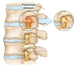
Precise decompression procedures to relieve pressure without excessive bone removal.

Spine Trauma Management

Stabilisation for fractures, dislocations and spinal cord injury with timely neuro-protective care.

Deformity Correction

Surgical correction for scoliosis, kyphosis and postural deformities with long-term follow-up.

Why Choose Dr. Nagesh Chandra?

Minimally Invasive Approach

Uses incision-sparing procedures to reduce blood loss, pain and recovery times.

Precision Imaging-Based Planning

Combines MRI, CT and neuro-navigation for highly targeted surgical intervention.

Outcome-Driven Treatment

Focus on restoring mobility, reducing nerve pain and preventing long-term spine dysfunction.

Multidisciplinary Coordination

Works with neurology, rehab, radiology and pain specialists for holistic care.

Transparent Guidance

Explains spine conditions, surgical need and expected outcomes clearly to build patient trust.

Achievements & Recognitions

High-Volume MIS Surgeon

Performed numerous minimally invasive spine procedures with consistent postoperative outcomes.

Key Contribution to Spine Protocols

Helped develop hospital guidelines for early mobilization after spine surgery.

Faculty at Ortho-Spine Workshops

Invited speaker and trainer for cadaveric spine dissection and MIS instrumentation courses.

Clinical Research Participation

Part of observational studies on degenerative spine disease and surgical recovery metrics.

ER Spine Response Leadership

Led multiple emergency cases involving complex spine trauma management.

Awards

Orthopedic Excellence Award

Awarded for consistent clinical results in complex spine surgery cases.

Hospital Service Appreciation

Recognised for patient-care quality and multidisciplinary coordination.

Teaching Excellence

Awarded for training junior orthopedic and spine fellows.

Quality & Safety Recognition

Honoured for contributing to safer operation theatre protocols.

Peer Recognition

Frequently acknowledged by colleagues for dependable surgical judgment.

Professional Memberships

Association of Spine Surgeons of India (ASSI)

Active participant in national spine meetings and conferences.

Indian Orthopaedic Association (IOA)

Member contributing to clinical discussions and continuous medical education.

Minimally Invasive Spine Society

Attends MIS technique-specific workshops regularly.

Hospital Surgical Board

Part of surgical planning committees and quality audit panels.

Regional Trauma Network

Collaborates with trauma centres for coordinated spine-injury care.

Trust & Editorial Policy

This content adheres to the CureU Editorial Guidelines, ensuring evidence-based accuracy and regular updates.

Let Us Help You

Free Expert Consultation

    By submitting the form I agree to the Terms of Use and Privacy Policy of CureU Healthcare.

    Treatments under Neurology and Spine

    Craniofacial Reconstruction Surgery

    Neurology and Spine

    Craniofacial Reconstruction Surgery

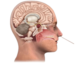
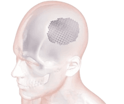
    Craniofacial surgery is used to treat issues with the head, skull, face, and neck. Specifically, the treatment involves surgical procedures and the supervision of skilled surgeons. Craniofacial reconstruction surgery reconstructs the damaged bone and tissue; as a result, this helps in improving the appearance of disfigured or abnormal shapes of the face and head. For instance, children with abnormal facial structures benefit from early surgery, which is advantageous in minimizing the impact of these conditions on growth, development, and function. -br There are generally two groups of people who may need craniofacial surgery. First, the first group of people are those who are born with abnormalities of the bones, muscles, and other tissues of the skull and face. This condition is known as congenital abnormalities. -br On the other hand, the second group includes people who develop irregularities in the skull and face later in life. This can occur due to diseases and trauma. This condition is known as acquired abnormalities.

    Chat with Us
    Discectomy

    Neurology and Spine

    Discectomy

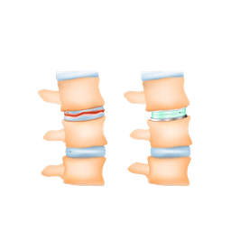
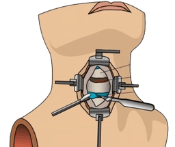
    A discectomy is a surgical procedure to remove some part or complete part of an intervertebral disk in the spine. To explain further, intervertebral disks are like flat, round cushions that are placed between the vertebrae (bones) in the spine and work as spine shock absorbers. Each disk has a soft, gel-like centre called nucleus pulposus, surrounded by a flexible outer ring called annulus. Importantly, intervertebral disks are under regular pressure. As a result, a disk can tear, allowing some of the nucleus gel substances that come out through leaking. This condition is known as a herniated disk, also known as bulging, slipped, or ruptured disk. Consequently, this is the main reason that leads to performing a discectomy. -br In most cases, herniated disks mostly affect the lower back called the lumbar spine, but in addition, they also affect the neck called cervical spine too. However, a rare place where herniated disks are seen is the middle back. -br A discectomy in India, also spelled as diskectomy.

    Chat with Us
    Meningioma

    Neurology and Spine

    Meningioma

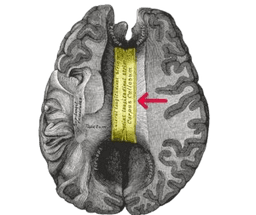
    Meninges are the layer of tissue that protects the brain and spinal cord. A meningioma is a brain tumor that develops in meninges. Meninges are made of three layer tissues that cover and protect the brain and spinal cord. Meningiomas start from cells made up of particular arachnoid cells which are found in a thin, web-like layer that covers the brain and spinal cord. This layer is one of those layers that protects and makes up the brain's covering protective system known as meninges. -br Meningiomas in India are not always cancerous (benign), although sometimes they can be cancerous and malignant. If a tumor is cancerous it means it's aggressive or can spread out in other tissues as well or can spread in other parts of the body. A benign tumor is the type of tumor that can't spread to other parts of the body. -br Meningiomas are mostly located near the top or outer curve of the brain. They also form at the base of the skull. Spinal meningiomas are very uncommon. -br Meningiomas develop slowly and inward. They do not show any symptoms before they are diagnosed. Even benign meningiomas can grow larger enough to be life threatening if they compress and affect nearby areas of the brain.

    Chat with Us
    Spina Bifida

    Neurology and Spine

    Spina Bifida

    Spina bifida is a medical condition of an unborn fetus that means incomplete development of the spine during the first month of pregnancy. Moreover, this is the most common inborn disorder that is mostly affected in the United States. The term spina bifida means split spine. -br Specifically, spina bifida occurs during the first 28 days of pregnancy, even before the pregnancy is confirmed by the women. The condition is a type of neural tube defect (NTD). -br In addition, in some uncommon cases, the spina bifida in India is very serious. The condition varies widely in degree. Generally, most of the cases are mild that have no symptoms and even can be treated without any serious treatment. -br However, in more uncommon cases of such disorder, infants are born with open lesions on their spine that lead to damage to surrounding nerves and spinal cord. The opening can be treated through surgery, but the nerve damage can't be solved and results in permanent disability. Furthermore, spina bifida can take place anywhere in the back bone.

    Chat with Us
    Cerebral of Brain Aneurysm

    Neurology and Spine

    Cerebral of Brain Aneurysm

    Cerebral aneurysm, also known as brain aneurysm, is a small and thin spot on the artery in the brain that bulges out and fills with blood. As a result, the bulging aneurysm leads to force or puts pressure on the nerves or brain tissue. This can also result in a burst, spilling blood surrounding tissue, known as hemorrhage. Consequently, a ruptured aneurysm can be a life-threatening condition, such as hemorrhagic stroke, brain damage, coma, or sometimes death.-br However, some cerebral aneurysms do not bleed, and are very small in size or cause other complications. These types of aneurysms are diagnosed while imaging tests for different medical problems. Cerebral aneurysms can take place anywhere in the brain, but most develop in major arteries along the base of the skull.-br Cerebral of brain aneurysms can develop at any age and anywhere in the brain. They are, however, most common in adults between the ages of 30 and 60 years, and are more common in females than males. Additionally, some patients are at high risk due to inherited disorders.

    Chat with Us
    Deep Brain Stimulation for Parkinson's Disease

    Neurology and Spine

    Deep Brain Stimulation for Parkinson's Disease

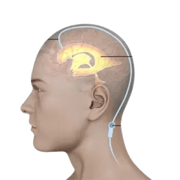
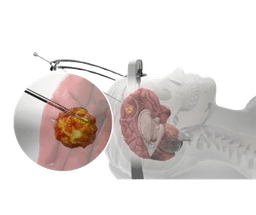
    Deep brain stimulation is a technique and a medical procedure that proceeds with a mild electric current delivered to the specific part of the brain. This means that this electricity in the current stimulates the brain cells in the area that treats several conditions of the brain and body. Specifically, the current travels through one wire or more wires and reaches to the brain connected with a small device implanted underneath the skin near the collarbone. -br Currently, deep brain stimulation is used for conditions like Parkinson's disease and epilepsy, although research is still on if it can be helpful for other conditions too. -br In this procedure, the treatment involves an implanted device that delivers electrical current directly to the brain areas.

    Chat with Us
    Early Onset Scoliosis Surgery

    Neurology and Spine

    Early Onset Scoliosis Surgery

    Early onset scoliosis involves the development of abnormal lateral curvature of the spine before the age of 10. The condition occurs at congenital or acquired different challenges because the spine is still in a growing stage. The majority of the children are diagnosed with this condition between the ages of 10 to 15 with adolescent idiopathic scoliosis. Identifying idiopathic scoliosis at birth or in early childhood is rare. The type of scoliosis is categorized by age at onset of diagnosis:

    Chat with Us
    Neurolysis Procedure

    Neurology and Spine

    Neurolysis Procedure

    Neurolysis is a procedure that consists of chemical injection that involves pain relief management. Generally, this is typically for nerve pain, cancer pain or visceral pain. Depending on the case, different types of chemical neurolysis procedure based on which nerve it's pointing out, such as celiac plexus neurolysis and intercostal nerve neurolysis. Typically, the injected chemical consists of alcohol, phenol or glycerol. -br To understand this, nerves are like the wires of the body that travels the electrical signal between the brain and the body. These electrical signals lead to feeling sensation like touch, pinch and pain and move the muscles. Therefore, the goal of neurolysis is to stop the targeted nerve from sending pain signals to the brain. As a result, if these nerves stop sending signals, the body won't be able to feel any sensation or do not respond accordingly. -br In general terms, intentional destruction of the pain management through the nerves is said to be as neurolysis in general ways. In other words, this neurolysis in India is a term used to describe the work of the nerve that passes the signals to the brain.

    Chat with Us

    Best Doctors for Neurology and Spine

    Dr. Amit Srivastava

    26+ years

    Dr. Amit Srivastava

    Neurology and Spine

    Profile
    Call Us
    Chat now

    How can I help you?