Overview
Dental bone grafting is a reconstructive procedure that restores bone volume and density in the jaw where tooth loss, periodontal disease, or trauma has caused deterioration.
The graft creates a scaffold that encourages the patient’s own bone to grow in and integrate, producing a stable foundation for implants, bridges, or to preserve facial contours.
Treatment is personalized: graft material may be autograft (your own bone), allograft (donor), xenograft (animal-derived), or synthetic, chosen to match the defect, healing timeline, and long-term goals.
With careful planning and follow-up, bone grafting reliably restores form and function so restorative dentistry has a predictable platform to succeed.
Overview And Clinical Background
Why Bone Volume Matters For Oral Health
Bone supports teeth, maintains facial shape, and anchors dental implants.
When a tooth is lost or periodontal disease progresses, the underlying bone resorbs progressively, creating defects that complicate later restorative work.
Bone grafting rebuilds these defects by providing a biologic scaffold that promotes new bone formation and restores structural integrity for implants or prosthetic teeth.
- Indication:: Deficient ridge height or width after tooth loss, extraction sockets needing preservation, bone loss from periodontal disease, or preparation for implant placement where primary stability is otherwise unattainable.
- Graft choices:: Autograft (patient bone), allograft (human donor bone), xenograft (processed animal bone), or synthetic substitutes—each with different remodeling profiles and indications that your surgeon will match to your needs.
- Biologic principle:: Graft material acts as scaffold (osteoconduction) and may also deliver cells or factors that recruit new bone-forming cells (osteoinduction) so native bone gradually replaces the graft.
Symptoms, Signs And Presentation
Patients present for bone grafting when insufficient bone prevents implant placement or when the jaw has collapsed after long-standing tooth loss.
Signs include narrow ridges, loose dentures that no longer fit, or visible facial volume loss that alters smile aesthetics.
Pain is not usually the presenting complaint; rather the functional or aesthetic limitations prompt evaluation.
- Common presentation:: Collapsed alveolar ridge after long-term tooth loss leading to unstable dentures, difficulty chewing, or poor esthetic support of lips and cheeks.
- Functional issue:: Inability to place an implant of appropriate length or diameter because of insufficient vertical or horizontal bone.
- Red flag:: Active infection at the proposed graft site (untreated periodontal or endodontic infection) which must be controlled before grafting to ensure predictable healing.
Diagnosis Methods And Investigations
Imaging And Preoperative Assessment
Thorough clinical exam and 3D imaging guide graft design and material selection.
Cone-beam CT (CBCT) is the cornerstone investigation, quantifying bone volume and showing critical anatomy such as the sinus floor and nerve canals.
Medical history, smoking status, metabolic disease, and medications are reviewed because systemic factors influence graft integration.
- CBCT imaging:: Provides cross-sectional views of ridge width, height, and bone quality so the surgeon can plan graft size, harvesting site (if needed), and implant positioning with millimetre precision.
- Clinical tests:: Intraoral examination, periodontal charting, and assessment of soft tissue quality to ensure a sealed, tension-free closure over the graft—critical for success.
- Medical review:: Blood tests or medication checks when indicated (e.g., bisphosphonates, poorly controlled diabetes) to mitigate healing risks and tailor perioperative care.
Treatment Options And Surgical Techniques
Techniques range from simple socket preservation at time of extraction to block grafts and guided bone regeneration for large defects.
Surgeons combine graft material with barrier membranes, growth factors, or platelet concentrates to enhance healing and reduce resorption.
Choice of technique balances defect size, desired timeline for implant placement, and patient-specific healing capacity.
- Socket preservation:: Grafting the extraction socket immediately after tooth removal to minimize ridge collapse and simplify later implant placement—often using particulate graft and a resorbable membrane.
- Ridge augmentation:: Onlay or block grafting for horizontal or vertical deficiencies where a cortical block (often from the patient’s own jaw) or stacked particulate graft rebuilds form prior to implant placement.
- Sinus lift and guided bone regeneration:: Elevating the sinus floor with graft material to gain vertical height for implants in the posterior maxilla, combined with membranes to direct bone formation and prevent soft tissue ingrowth.
Recovery, Risks And Prognosis
Healing typically spans months as the graft remodels into vital bone; initial soft tissue healing occurs in days to weeks.
Risks include infection, graft exposure, partial graft failure, or donor site morbidity when autograft is used.
Smoking, uncontrolled systemic disease, or poor oral hygiene increase failure risk; conversely, careful surgical technique, proper soft tissue closure, and adherence to postoperative instructions yield high success rates and predictable implant support.
- Typical timeline:: Soft tissue closure in 1–2 weeks; radiographic evidence of graft integration over 3–6 months before implant placement in most cases; some rapid protocols vary by graft type.
- Possible complications:: Infection, wound dehiscence exposing graft, numbness if nerves are involved during harvesting, or partial graft resorption requiring secondary grafting in rare cases.
- Long-term outlook:: When integrated, grafted bone provides stable implant support and preserves facial contours—long-term prognosis is excellent with maintenance and regular dental follow-up.
Why Choose Us
CureU Healthcare combines digital planning, 3D imaging, and experienced oral surgeons to deliver predictable bone grafting outcomes.
We personalize graft selection, minimize donor site morbidity, and integrate minimally invasive harvesting when possible.
Our team ensures clear communication, staged treatment planning for timely implant rehabilitation, and detailed aftercare to maximize graft survival and patient comfort.
- Digital precision:: CBCT-guided planning and surgical guides enable accurate graft sizing and implant placement, reducing surprises and speeding recovery.
- Multidisciplinary care:: Coordination between surgeons, restorative dentists, and hygienists ensures both biological success and esthetic restoration.
- Aftercare support:: Structured postoperative protocols, clear instructions, and timely review visits help detect and manage complications early for durable results.
Conclusion
Dental bone grafting rebuilds the foundation needed for long-term oral rehabilitation—restoring function, supporting implants, and preserving facial form.
With proper assessment, skilled execution, and dedicated follow-up at CureU Healthcare, most patients achieve predictable bone regeneration and successful implant-supported restorations.

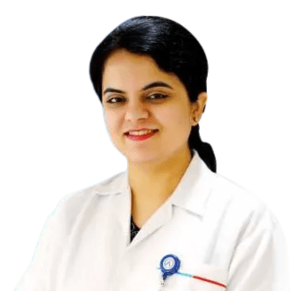
B.U.M.S, M.D. (NAT) | Reg No. 9153 | Ex. Secretary & Treasurer, N.I.M.A.
Qualified healthcare professional in Unani and Naturopathic medicine with deep expertise in traditional healthcare systems, patient-centered clinical care, and medical content review.